
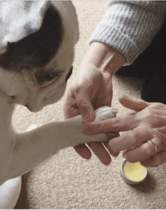
In mild cases, the optimal treatment option may be observed without treatment. In dogs with marked hyperkeratosis and a significant excess of keratin, it is removed by means of scissors or a blade. After appropriate instructions, pet owners are able to carry out this procedure at home, and this can serve as the only correction method. In most cases, trimming it is applied only at the beginning of treatment with the subsequent removal of excess keratin through hydration and application of moisturizing and emollients. Here are the details information about hyperkeratosis dogs .

Local treatment, as mentioned above, begins with cutting off excess keratin, and then a moisturizing compress is applied to the lesion for 5-10 minutes, followed by the application of keratolytic agents. When cracking, an ointment with antibiotics and corticosteroids is used. As a rule, the duration of the initial treatment is 7-10 days, during which time the affected surface returns to a state close to normal, after which the treatment either stops for a while or continues with a reduced frequency 1-2 times a week. In each case, the treatment is selected based on the state of the lesions and the wishes of the owner of the animal.
Parasympathetic hyperkeratosis of the nose of dogs
Parasympathetic dysfunction can lead to loss of function of the normal gland of the nose, which is localized in the lateral part of the nasal mucosa. This gland has parasympathetic innervation from the facial nerve.
Dermatosis can remain stable, weaken and intensify or progressively deteriorate. Lesions are usually limited to the nose, but mild scaly and crusted formation can be observed on the hairy part of the nose. Sick dogs are usually evaluated for the presence of dry keratoconjunctivitis and otitis.
Treatment and prognosis
The intensity of therapy depends on the severity of the lesions. The treatment used for idiopathic hyperkeratosis of the nose should be used. The forecast is good. The development of corns is often observed in large short-haired breeds that sleep on a hard surface. The places of occurrence are usually the outer side of the elbow and hock joints. They may also develop on the sternum of dogs, with a deep chest, or dogs with short legs, in which the sternum constantly contacts objects, such as stairs. Corns look like a bald area with a dense, cornified skin, with a grey surface. Often in short-haired breeds, hair is compressed within the follicles and corns. Ingrown hair, fistulas, secondary infection may occur.
The diagnosis is usually based on history and clinical manifestations. Further research may be needed to identify the underlying diseases in cases of infection, not responding to antibiotics or non-healing wounds. For uninfected lesions, monitoring without treatment is advisable. If the lesion is again infected, prescribe a long-term systemic antibiotic therapy at least 4-6 weeks. Alternatively, topical treatment may be used.
First, you need to remove the “ingrown hair” from the corn because they will lead to furunculosis and infection. Perform peeling of hair often. When using adhesive tape, only ingrown hairs are removed, leaving healthy and active intact. Bedding and other areas for sleeping and rest should be soft, and bandaging should be used to prevent injury to the affected area. Moisturizers, antibiotic ointments should be used for the local treatment of the damaged area every 12-24 hours to soften the skin. Surgical removal is usually not recommended, as a possible complication is the divergence of the wound edges.